The Role of Altered Brain Pathways
Could an internal breakdown in communication be driving the external breakdown in communication?
The complexity of the human brain lies not only in the sheer number of neurons (an estimated 86 million) but also in the vast network of connections, or synapses, between them. The brain communicates with other parts of the body through the transmission of signals along specialized pathways, allowing the brain to coordinate and control various functions throughout the body, including voluntary and involuntary movements, sensory perception, and the regulation of physiological processes.
This intricate interplay of neurons that are carrying messages enables the brain to perform its diverse functions. However, communication can break down if there is a disruption anywhere along the pathway. Dystonia is considered a network or circuit disorder involving abnormal interactions in regions of the brain that coordinate and control movement. In spasmodic dysphonia (SD), a form of focal dystonia, signals are misfired to the muscles of the larynx, causing those muscles to adduct (close) or abduct (open) unexpectedly, affecting the sound of a person’s voice.
The basal ganglia-cortical network, which includes interconnected brain regions responsible for planning and executing voluntary movements, is thought to be affected in dystonia. There is a breakdown in sensory input and motor output integration causing uncontrollable movements. Here we explore some of the areas that are part of this network and the role they play in normal movement.
Basal Ganglia
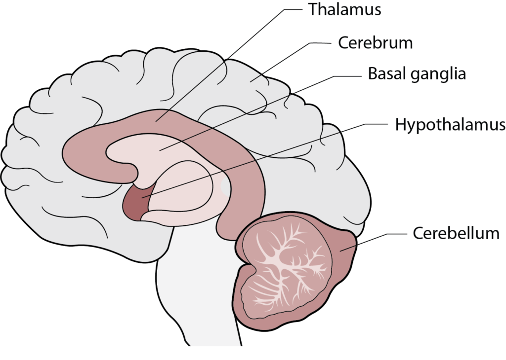
WHAT | The basal ganglia is a collection of neurons that form specific brain regions that play a crucial role in motor control and is involved in various functions, including the regulation of voluntary movements, motor planning, and coordination. These structures are interconnected in a complex circuitry that involves both direct and indirect pathways. The basal ganglia work in collaboration with other brain regions, such as the thalamus, cerebellum and cerebral cortex, to regulate motor functions and contribute to motor learning.
WHERE | The basal ganglia are a group of neurons located deep within the brain, near the base of the forebrain. They are situated on both sides of the brain, with each side containing its own set of basal ganglia structures, the major components being the striatum, globus pallidus, subthalamic nucleus, and substantia nigra.
Cerebellum
WHAT | The cerebellum is a large, separate brain structure that contributes to the smooth execution of movements, maintenance of balance, and the learning of motor skills. Its intricate connections with other parts of the nervous system make it a central player in motor control and coordination.
WHERE | The cerebellum is located in the back of the brain and directly connects to the spinal cord, brain stem, and thalamus. Via these connections, the cerebellum plays a fundamental role in coordinating voluntary movements and maintaining balance and posture.
Thalamus
WHAT | The thalamus is an integral part of the motor control circuitry in the brain. While it doesn’t directly generate movements, its role in relaying sensory information and integrating motor signals from different brain regions is crucial for the smooth execution and coordination of voluntary movements.
WHERE | The thalamus is found above the brainstem and cerebellum, and below the cerebral cortex and basal ganglia. It is composed of two symmetrical halves, or hemispheres, each containing several nuclei that relay motor and sensory signals to different regions of the body or cerebral cortex, respectively.
Cerebral Cortex
WHAT | The cerebral cortex is intricately involved in the control of high-level voluntary movement, with different regions contributing to various aspects of motor planning and execution. The cerebral cortex integrates sensory information with intended motor plans to execute intended movement.
WHERE | The cerebral cortex is the outermost layer of the brain, often referred to as the “gray matter” of the brain due to its gray appearance. It makes up the majority of the brain’s mass and is divided into four main lobes.
Somatosensory System
WHAT | The somatosensory system monitors the environment via sensory receptors, nerves, and brain regions. This system is responsible for detecting, processing, and interpreting sensations related to touch, pressure, temperature, pain, and body position (proprioception). It provides essential feedback for spatial awareness and motor control and optimizes interactions with the environment. Integration of somatosensory information occurs at various levels of the nervous system, including the basal ganglia, cerebellum, thalamus, and cerebral cortex, allowing for a comprehensive understanding of the body’s external and internal states.
WHERE | The primary components of the somatosensory system are distributed throughout the body, including sensory receptors, peripheral nerves, spinal cord, brainstem, thalamus, and cerebral cortex.
DYSTONIA AND ITS RELATIONSHIP TO THE NERVOUS SYSTEM
Neurotransmitter Imbalance
Disruptions in the balance of chemical messengers in the brain (neurotransmitters) occur in dystonia. The basal ganglia-cortical network relies on precise neurotransmitter signaling for smooth motor control and imbalances can result in increased activity of motor circuits, contributing to dystonic movements. Specific to dystonia, the most commonly affected neurotransmitter systems include dopamine, GABA, and acetylcholine. Disruptions in neurotransmitter balance may contribute to abnormal brain signals that affect precise control of laryngeal muscles. The exact mechanisms and their relationships are complex and not fully understood.
Genetics
While many cases of dystonia are considered sporadic and occur without a clear family history, there are genetic factors that contribute to certain forms. Some individuals may have a genetic predisposition to dystonia, meaning that their genetic makeup increases their likelihood of developing dystonia. Currently, there are no identified genes that directly cause spasmodic dysphonia.
Environmental Factors
Environmental factors, such as trauma, stress, infections, or illness, may have a role in the onset of spasmodic dysphonia. It is unclear whether these things directly trigger spasmodic dysphonia or if they exacerbate pre-existing conditions. In addition, when combined with genetic susceptibility, environmental factors may contribute to the development of spasmodic dysphonia.
Abnormal Sensory-Motor Integration
Dystonia is associated with abnormal sensorimotor processing, affecting the integration of sensory information with motor commands. Continuous feedback loops between the somatosensory system and motor areas contribute to adjusting movements in real-time based on sensory input. In dystonia, the brain struggles to interpret feedback from the body correctly, leading to abnormal muscle contractions.
The somatosensory system for voice involves sensory receptors in the vocal tract, larynx, and other related structures that provide feedback to the brain about the position, movement, and tension of the vocal apparatus. When this system is disrupted, it can impact speech production and the perception of one’s own voice.
Abnormal Neuroplasticity
Neuroplasticity refers to the brain’s capacity to change and reorganize itself by altering existing or new connections throughout life. Mechanisms of neuroplasticity are being actively investigated, and much is being learned about changes in the brain that cause this. For example, connections between nerve cells can be modified to convey more strong or weak messages. Additionally, nerve processes can grow or shrink. The location in the brain where these alterations occur remains unclear and is being investigated. In dystonia, the role of neuroplasticity is not fully understood, but there is evidence to suggest that unfavorable neuroplasticity changes affect the brain’s motor networks. It may be possible to reverse these changes, but a robust method is not yet available. More understanding of the cause is needed before these changes can be reversed.
Network Dysfunction
It has long been believed that connections between the basal ganglia and cerebral cortex form a connection (network) disrupted in people with dystonia. There is growing evidence that disruption of additional brain connections may contribute to dystonia, including those that involve the cerebellum, thalamus, and cerebral cortex. These complex interactions lead to the consideration that dystonia results from the disruption of signals in a brain network that controls movement. This perspective underscores the complexity of dystonia and the need for a comprehensive understanding of the interconnected brain regions involved in motor control.
Conclusion
Spasmodic dysphonia is a prime example of the intricate and interconnected nature of the human brain. When these connections are disrupted, as seen in SD, the effects ripple outward, significantly impacting the voice and complicating daily life for those affected. Emerging research reveals that the causes of these disruptions are multifaceted, including neurotransmitter imbalances, abnormal sensory-motor integration, and unfavorable neuroplasticity. This complexity underscores that SD is not merely a local issue of misfiring signals in the larynx but a disorder deeply rooted in the interplay of brain networks responsible for movement control. Understanding these dynamics is essential for advancing diagnostic techniques, refining treatment options, and paving the way toward finding a cure. Environmental factors and genetic predispositions further add layers of complexity but also open the door to more personalized and effective interventions. By restoring communication within the brain’s motor circuits, researchers may be able to reverse unfavorable neuroplasticity and recalibrate disrupted pathways. Through continued exploration and targeted research, we move closer to not only restoring voices but also enhancing our broader understanding of brain function and its role in regulating communication and movement.
NETWORK VS. CIRCUIT DISORDER
The terms “network disorder” and “circuit disorder” are often used interchangeably in the context of neuroscience and neurology. Both refer to conditions that involve abnormalities or dysfunction of neural networks or circuits within the brain. However, there can be subtle distinctions in how these terms are used, depending on context.
“Network disorder” typically implies a broader perspective, referring to abnormalities or dysregulation in the communication and interactions among various brain regions. It emphasizes the interconnectedness of the different brain structures and their roles in supporting specific functions or behaviors. Examples of network disorders include conditions resulting from disrupted communication between brain regions, resulting in problems with cognitive, sensory, or motor function.
“Circuit disorder” highlights specific brain circuits or connections that are dysfunctional. It often implies a more focused consideration of particular brain circuits involved in a specific function or behavior. Examples of circuit disorders include conditions where specific pathways, such as those in the basal ganglia-cortical circuit, are implicated in the cause of a disorder.
In essence, both terms emphasize the idea that neurological disorders may arise from disturbances in any part of intricate connections and interactions between different brain regions that define networks or circuits.


